
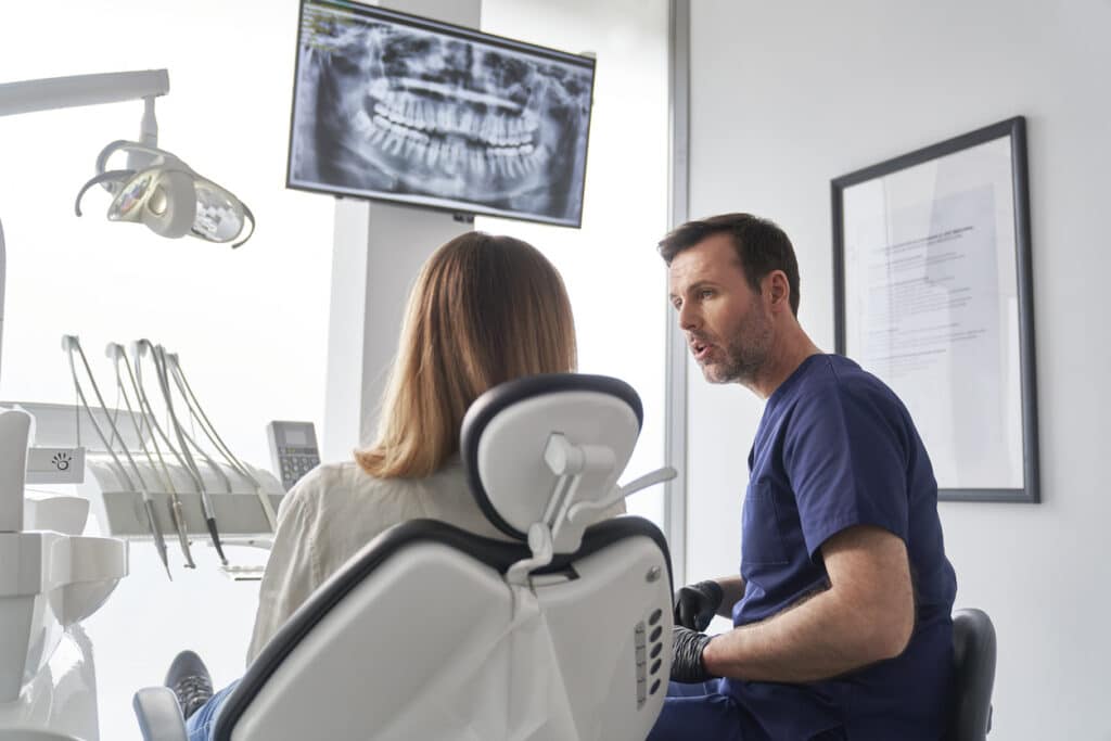
Dental X-rays at Cary Towne Dental are advanced diagnostic imaging tools used to detect hidden decay, bone loss, and oral health issues before they become severe. These safe, routine scans are performed using low-dose digital radiography and strict guidelines to minimize radiation exposure while maximizing preventive care for North Carolina patients. Notably, clinical literature supports that wireless and wired sensors for X-rays significantly reduce the amount of radiation administered to the patient compared to conventional X-rays [1] (Ardila et al., 2024).
The Environmental Advantage: A Greener Choice for Dental Care
Medical evidence indicates that Hazards to health from exposure to chemicals in the automatic processing of photographic film are well documented [2] (Hewitt, 1993). Older analog processes use developer and fixer solutions that contain dangerous substances such as silver halide. Digital X-rays eliminate this chemical waste completely. When you choose digital imaging, you actively help keep the local Wake County water supply clean. There is zero need for disposable plastic film packets, harmful lead foils, or harsh liquid processing solutions. For the health-conscious patient, this is a clean, sustainable, and forward-thinking approach to preventive dental care.
Beyond the Surface: What Digital X-Rays Actually Detect
A standard visual exam only tells half the story. No matter how thoroughly you brush and floss, your dentist cannot see between your tightly spaced teeth or deep beneath your gum line.
Digital Sensors: Digital sensors make a high-definition map of your oral health. They spot micro-cavities lurking in the tight spaces that your toothbrush bristles cannot reach. They also show early signs of bone loss from gum disease, the position of impacted wisdom teeth, and locate hidden root infections. This way, you catch these hidden issues early on and avoid expensive root canals or painful extractions later.
Pediatric Dental Safety: Gentle Imaging for Younger Patients
Digital dental X-rays are perfectly engineered for growing, sensitive smiles. Medical literature indicates that children and adolescents are particularly sensitive to ionizing X-rays [3] (Hennig et al., 2023). This is why we utilize special, small-scale pediatric sensors specifically designed with rounded edges to fit comfortably inside a child’s mouth. Because the image capture happens in a fraction of a second, even the most energetic toddlers can get clear diagnostic results without needing to sit perfectly still for long periods. This fast, gentle process helps alleviate childhood dental anxiety and builds a strong foundation for lifelong oral health.
Traditional Film vs. Digital Dental X-Rays
When choosing a dental home, it helps to understand the tools they use. Here is how modern digital technology compares to older traditional methods.
Feature | Traditional Film X-Rays | Advanced Digital X-Rays |
Radiation Exposure | Standard baseline | Up to 90% less radiation |
Processing Time | 10–15 minutes | Instant (Under 3 seconds) |
Maintenance Level | Requires toxic developing chemicals | Zero chemicals; clean digital processing |
Storage & Durability | Physical files that degrade over time | Cloud-stored; easily shared with specialists |
Cost Profile | Ongoing material costs (film/chemicals) | Lower long-term costs; usually covered by insurance |
Seamless Integration: Planning Your Future Smile
Digital X-rays do more than just find cavities; they act as the precise blueprint for your future treatments. If you are a career-focused professional looking to upgrade your smile, this technology accelerates your results. These high-definition digital files provide precise measurements of the density of your jawbone if you are considering undergoing cosmetic enhancements or restorative work such as dental implants. Also, these 2D images can be combined with sophisticated 3D scanners to plan the clear aligner treatments with accurate precision. This is a data-driven, high-accuracy plan that means fewer trips back to our Cary office and results you can count on for your busy schedule.
Co-Diagnosis: Building Trust Through Visual Transparency
Dental anxiety often stems from a simple fear of the unknown. When a dentist tells you that you need a filling or a crown, you deserve to understand exactly why. Digital X-rays remove the mystery from your diagnosis. Because the crisp images display instantly on a monitor right beside your dental chair, you and your dentist look at the exact same screen. Your dentist can zoom in, adjust the color contrast, and point directly to the area of concern. This collaborative, transparent approach puts you firmly in control of your healthcare decisions.
FAQS
1. Do I really need bite-wing X-rays every year?
Not necessarily. While annual bite-wings are standard for patients with a history of decay, your dentist may extend this timeframe to 18 or 24 months if you maintain excellent oral hygiene and show a low risk for cavities.
2. Are dental X-rays safe during pregnancy?
Yes. According to the American Dental Association, dental X-rays are safe during pregnancy with the use of a protective lead apron and thyroid collar. However, we often postpone routine X-rays until after delivery for your complete peace of mind, only taking them during pregnancy if an emergency arises.
3. How does Cary Towne Dental protect patients from radiation?
We strictly follow the ALARA principle—As Low As Reasonably Achievable. We use highly sensitive digital sensors that require a fraction of standard radiation, employ lead aprons with thyroid collars, and only prescribe X-rays based on your individualized health needs.
References
[1] Ardila, C. M., Vivares-Builes, A. M., & Pineda-Vélez, E. (2024). Image Quality, Radiation Dose, and Patient Comfort Associated with Wireless Sensors in Digital Radiography: A Systematic Review. Dentistry journal, 12(8), 267. https://doi.org/10.3390/dj12080267
[2] Hewitt PJ. Occupational health problems in processing of X-ray photographic films. Ann Occup Hyg. 1993;37(3):287-295. doi:10.1093/annhyg/37.3.287
[3] Hennig, C. L., Schüler, I. M., Scherbaum, R., Buschek, R., Scheithauer, M., Jacobs, C., & Mentzel, H. J. (2023). Frequency of Dental X-ray Diagnostics in Children and Adolescents: What Is the Radiation Exposure?. Diagnostics (Basel, Switzerland), 13(3), 394. https://doi.org/10.3390/diagnostics13030394