
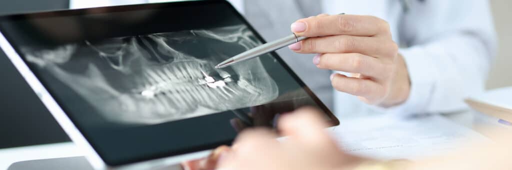
Digital X-rays are a low-radiation diagnostic imaging tool that captures instant, high-definition pictures of your teeth and jawbone. Additionally, medical evidence shows that Digitized images make it easier to implement a picture archiving and communication system [1] (Almanei et al., 2017). This modern technology allows dentists to detect cavities, bone loss, and root infections early while keeping your appointment fast and highly accurate.
What are digital X-rays?
Modern digital X-rays at Cary Towne Dental offer a seamless, patient-focused diagnostic process. Medical evidence shows that digital intraoral radiographs provide excellent spatial resolution for detecting caries and periodontal bone loss [2] (Oliveira, 2025). Instead of film, your dental assistant places a small electronic sensor in your mouth. This sensor connects to our secure computer system. When the image is captured, it does not require the toxic chemicals typically used in a darkroom process. The data is sent directly to the monitor in the room, showing your tooth roots and jawbone. This means you and the dentist see the exact same data. This transparent, co-diagnosis approach removes the mystery from your dental care, allowing you to clearly see and understand the status of your oral health.
Fast-track process for patients who are busy
For busy professionals working in the Triangle, your time is valuable. Our modern dental workflow is engineered for efficiency. Clinical literature supports that digital dental X-ray images are an efficient and accurate way to conduct auxiliary diagnoses of dental diseases [3] (Liu et al., 2023). First, you wear a protective lead apron for safety. Next, the hygienist positions the sensor within your mouth. Image capture takes only a fraction of a second. Within three seconds, the image appears on the monitor. A full routine set of diagnostic X-rays is typically completed in less than 5 minutes. Because our dentists can instantly enhance the contrast and accurately measure bone density, your diagnostic review is precise. We can catch early signs of decay when they are affordable to fix.
Comparing digital imaging options
Diagnostic Imaging Type | Clinical Focus & Purpose | Cost Predictability | Maintenance Level (Frequency) |
Routine Bitewing X-Rays | Detects cavities hidden between tightly spaced back teeth. | Low (Often covered 100% by standard PPO insurance). | Routine (Typically taken every 12 to 24 months). |
Panoramic Digital Scan | Captures a wide, 2D view of your entire mouth, jaw joints, and impacted wisdom teeth. | Moderate (Covered by most plans every few years). | Low (Taken every 3 to 5 years for a broad health baseline). |
Periapical (PA) X-Ray | Focuses on the entire length of a single tooth, from the visible crown to the tip of the root. | Low. | As needed (Used specifically to diagnose a localized toothache or infection). |
How digital sensors work and capture important oral health information
Today, the digital sensor uses millions of microscopic electronic pixels to measure the exact density of your oral tissues. When the gentle energy passes through your mouth, dense materials like healthy enamel and jawbone register as bright white. Softer areas, such as hidden decay or an active root infection, show up as dark spots. This direct data transfer creates a crisp, high-resolution map of your mouth on our computer screen in under three seconds.
Discover bone loss before it has the ability to spread
Gum disease is a notoriously silent condition. You often will not feel any pain as harmful bacteria slowly eat away at the bone supporting your teeth. A standard visual exam only lets Dr. Prahalad see the pink gum tissue above the surface. High-definition digital X-rays look past the gums to reveal the exact height and density of your jawbone. By capturing these images during your routine visits, we can spot the earliest warning signs of bone degradation. Catching this damage early allows us to stop the infection quickly, keeping your natural teeth firmly in place and saving you from complex oral surgery.
FAQS
1. Will I gag during digital dental X-rays?
No, modern technology has made this process much more comfortable. Digital sensors are significantly smaller and thinner than old cardboard film packets. They have smooth, rounded edges that fit the natural curves of your mouth, drastically reducing the gag reflex and soft-tissue pinching.
2. How long do digital X-rays add to a routine dental checkup?
They add almost no time at all. Capturing a standard set of digital bitewing images takes less than five minutes. Because the images render on our screens instantly, you avoid the 10-to-15 minute wait times associated with outdated chemical film processing.
3. Can I transfer my digital X-rays to a specialist in Wake County easily?
Yes. Because your diagnostic images are securely stored as high-definition digital files, we can email them directly to an oral surgeon or orthodontist using strict, HIPAA-compliant encryption. You never have to worry about carrying physical files across town.
References
[1] Almanei, K., Alsulaimani, R., Alfadda, S., Albabtain, S., & Alsulaimani, R. (2017). Digitally Scanned Radiographs versus Conventional Films for Determining Clarity of Periapical Lesions and Quality of Root Canal Treatment. TheScientificWorldJournal, 2017, 2427060. https://doi.org/10.1155/2017/2427060
[2] Oliveira M. L. (2025). Digital Dental Radiology and Diagnostics-From 2D to 3D. Australian dental journal, 70 Suppl 1(Suppl 1), S50–S66. https://doi.org/10.1111/adj.70024
[3] Liu, F., Gao, L., Wan, J., Lyu, Z. L., Huang, Y. Y., Liu, C., & Han, M. (2023). Recognition of Digital Dental X-ray Images Using a Convolutional Neural Network. Journal of digital imaging, 36(1), 73–79. https://doi.org/10.1007/s10278-022-00694-9